File:PMC4785757 JGID-8-51-g002.png
Jump to navigation
Jump to search
PMC4785757_JGID-8-51-g002.png (512 × 523 pixels, file size: 223 KB, MIME type: image/png)
File history
Click on a date/time to view the file as it appeared at that time.
| Date/Time | Thumbnail | Dimensions | User | Comment | |
|---|---|---|---|---|---|
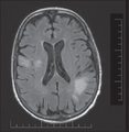
| current | 23:53, 3 February 2022 |  | 512 × 523 (223 KB) | Ozzie10aaaa | Author:Panchabhai TS, Choudhary C, Isada C, Folch E, Mehta AC,John and Doris Norton Thoracic Institute, St. Joseph's Hospital and Medical Center (Openi/National Library of Medicine) Source:https://openi.nlm.nih.gov/detailedresult?img=PMC4785757_JGID-8-51-g002&query=&req=4 Description:F2: Axial T2-weighted, fluid-attenuated inversion recovery magnetic resonance image shows progressive multifocal leukoencephalopathy with a high signal intensity lesion involving the white matter of the dorsal ri... |
File usage
The following page uses this file:
