File:PMC4750013 gr1.png
Jump to navigation
Jump to search
No higher resolution available.
PMC4750013_gr1.png (512 × 302 pixels, file size: 418 KB, MIME type: image/png)
File history
Click on a date/time to view the file as it appeared at that time.
| Date/Time | Thumbnail | Dimensions | User | Comment | |
|---|---|---|---|---|---|
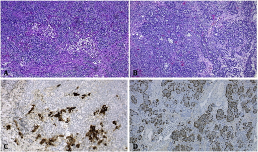
| current | 21:16, 13 April 2022 | | 512 × 302 (418 KB) | Ozzie10aaaa | Author:Liggins CA, Ma LT, Schlumbrecht MP,Department of Obstetrics and Gynecology at Banner University Medical Center (Openi/National Library of Medicine) Source:https://openi.nlm.nih.gov/detailedresult?img=PMC4750013_gr1&query=Sertoli%E2%80%93Leydig%20cell%20tumour&it=xg&req=4&npos=2 Description:f0005: Immunohistochemical staining of Sertoli–Leydig cell tumor. (A): Sertoli–Leydig cell tumor, intermediate grade with hepatoid differentiation. (B): Area of carcinoid tumor. (C): Immunohistochemi... |
File usage
There are no pages that use this file.