File:PMC4739221 CRIOT2016-3642735.001.png
Jump to navigation
Jump to search
No higher resolution available.
PMC4739221_CRIOT2016-3642735.001.png (512 × 375 pixels, file size: 336 KB, MIME type: image/png)
File history
Click on a date/time to view the file as it appeared at that time.
| Date/Time | Thumbnail | Dimensions | User | Comment | |
|---|---|---|---|---|---|
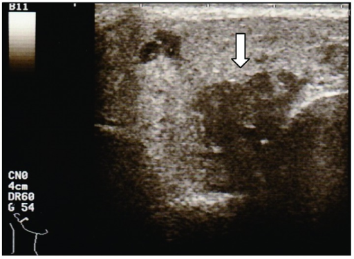
| current | 23:10, 16 March 2022 | | 512 × 375 (336 KB) | Ozzie10aaaa | Author:Fujiwara K, Furuta Y, Fukuda S, Department of Otolaryngology, Head and Neck Surgery, Graduate School of Medicine, Hokkaido University(Openi/National Library of medicine) Source:https://openi.nlm.nih.gov/detailedresult?img=PMC4739221_CRIOT2016-3642735.001&query=Heerfordt%20syndrome&it=xg&req=4&npos=2 Description:fig1: Ultrasound image for Case 1, showing the enlarged right parotid gland and interspersed hypoechoic areas (arrow). |
File usage
There are no pages that use this file.