File:PMC4706576 247 2015 3444 Fig1 HTML.png

From WikiProjectMed
Jump to navigation Jump to search

PMC4706576_247_2015_3444_Fig1_HTML.png(512 × 181 pixels, file size: 76 KB, MIME type: image/png)

This file is from a shared repository and may be used by other projects. The description on its file description page there is shown below.

Summary

Description
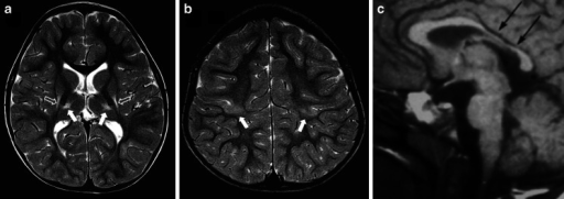
English⧼Colon⧽ Fig1: MR images in a 6-year old boy with dyskinetic cerebral palsy who has the typical neuroimaging findings of acute profound hypoxic–ischaemic brain injury. a, b Axial T2-weighted MR images at the level of the basal ganglia (a) and peri-rolandic cortex (b) show gliosis and reduction in volume of the putamina (open arrows), thalami and paracentral lobules (solid arrows). c A mid-sagittal T1-weighted MR image shows a focal reduction in thickness in the posterior part of the body of the corpus callosum (arrows)
Date
Source https://openi.nlm.nih.gov/detailedresult?img=PMC4706576_247_2015_3444_Fig1_HTML&query=Dyskinetic%20cerebral%20palsy&it=xg&req=4&npos=3
Author Stivaros SM, Radon MR, Mileva R, Connolly DJ, Cowell PE, Hoggard N, Wright NB, Tang V, Gledson A, Batty R, Keane JA, Griffiths PD

Licensing

{{subst:Custom license marker added by UW}} https://creativecommons.org/licenses/by/4.0/ Attribution 4.0 International (CC BY 4.0)

This file was uploaded with UploadWizard.

File history

Click on a date/time to view the file as it appeared at that time.

Date/TimeThumbnailDimensionsUserComment
current21:44, 11 February 2023Thumbnail for version as of 21:44, 11 February 2023512 × 181 (76 KB)Ozzie10aaaaUploaded a work by Stivaros SM, Radon MR, Mileva R, Connolly DJ, Cowell PE, Hoggard N, Wright NB, Tang V, Gledson A, Batty R, Keane JA, Griffiths PD from https://openi.nlm.nih.gov/detailedresult?img=PMC4706576_247_2015_3444_Fig1_HTML&query=Dyskinetic%20cerebral%20palsy&it=xg&req=4&npos=3 with UploadWizard

There are no pages that use this file.