File:PMC4664510 opth-9-2195Fig1.png
Jump to navigation
Jump to search
No higher resolution available.
PMC4664510_opth-9-2195Fig1.png (512 × 213 pixels, file size: 237 KB, MIME type: image/png)
File history
Click on a date/time to view the file as it appeared at that time.
| Date/Time | Thumbnail | Dimensions | User | Comment | |
|---|---|---|---|---|---|
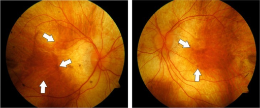
| current | 12:59, 12 October 2021 | | 512 × 213 (237 KB) | Ozzie10aaaa | Author:Zinkernagel MS, MacLaren RE. Department of Ophthalmology, Inselspital, Bern University Hospital, and University of Bern, Department of Clinical Research, Inselspital, Bern University Hospital, and University of Bern (Openi/National Library of Medicine) Source:https://openi.nlm.nih.gov/detailedresult?img=PMC4664510_opth-9-2195Fig1&query=Choroideremia&it=xg&req=4&npos=12 Description:f1-opth-9-2195: Fundus color photographs of the right and left eye of a patient with advanced choroideremi... |
File usage
The following page uses this file: