File:PMC4518818 cln-70-08-544-g003.png
Jump to navigation
Jump to search
No higher resolution available.
PMC4518818_cln-70-08-544-g003.png (512 × 344 pixels, file size: 355 KB, MIME type: image/png)
File history
Click on a date/time to view the file as it appeared at that time.
| Date/Time | Thumbnail | Dimensions | User | Comment | |
|---|---|---|---|---|---|
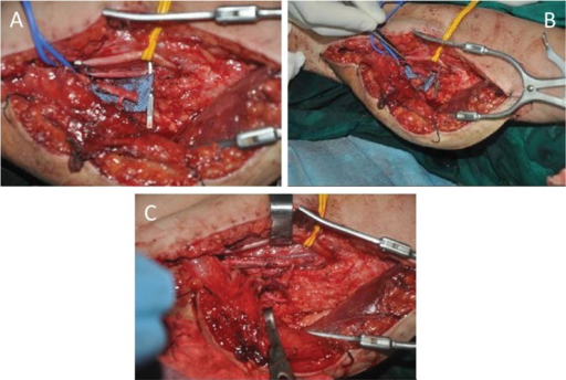
| current | 17:28, 7 September 2021 | | 512 × 344 (355 KB) | Ozzie10aaaa | Author:Hou Y, Yang J, Yang Y, Qin B, Fu G, Li X, Gu L, Liu X, Zhu Q, Qi J , Sun Yat-sen University (Openi/National Library of medicine) Source:https://openi.nlm.nih.gov/detailedresult?img=PMC4518818_cln-70-08-544-g003&query=Brachial%20plexus%20injury&it=xg&req=4&npos=42 Description:f3-cln_70p544: Flow-through anastomosis of the T-shaped pedicle. A) The diameter of the profunda femoris segment is obviously larger than that of the nutrient artery of the gracilis. B) The brachial artery was rese... |
File usage
The following page uses this file: