File:PMC4514984 12883 2015 369 Fig1 HTML.png
Jump to navigation
Jump to search
No higher resolution available.
PMC4514984_12883_2015_369_Fig1_HTML.png (472 × 493 pixels, file size: 216 KB, MIME type: image/png)
File history
Click on a date/time to view the file as it appeared at that time.
| Date/Time | Thumbnail | Dimensions | User | Comment | |
|---|---|---|---|---|---|
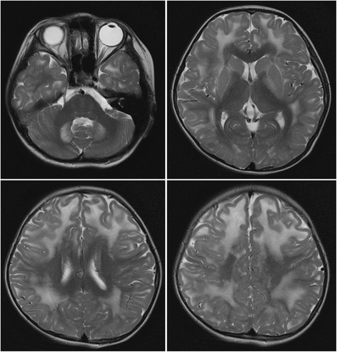
| current | 21:49, 10 December 2021 | | 472 × 493 (216 KB) | Ozzie10aaaa | Author:Tai H, Zhang Z , Department of Neurology, Beijing Tiantan Hospital, Capital Medical University(Openi/National Library of Medicine) Source:https://openi.nlm.nih.gov/detailedresult?img=PMC4514984_12883_2015_369_Fig1_HTML&query=2-hydroxyglutaric%20aciduria&it=xg&req=4&npos=3 Description:Fig1: The patient’s brain magnetic resonance image (MRI). Axial T2-weighted sequence of the brain MRI showed symmetrical subcortical white matter hyperintense involving bilateral dentate nucleus, internal... |
File usage
The following page uses this file: