File:PMC4435298 crj-01-131-g001.png
Jump to navigation
Jump to search
No higher resolution available.
PMC4435298_crj-01-131-g001.png (512 × 514 pixels, file size: 591 KB, MIME type: image/png)
File history
Click on a date/time to view the file as it appeared at that time.
| Date/Time | Thumbnail | Dimensions | User | Comment | |
|---|---|---|---|---|---|
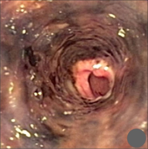
| current | 21:06, 8 December 2021 | | 512 × 514 (591 KB) | Ozzie10aaaa | Author:Abed J, Mankal P, Judeh H, Kim S, Department of Medicine, Icahn School of Medicine, Mount Sinai St. Luke's and Roosevelt Hospitals(Openi/National Library of medicine) Source:https://openi.nlm.nih.gov/detailedresult?img=PMC4435298_crj-01-131-g001&query=&req=4 Description:F1: Endoscopy performed at the time of admission shows an ulcerated and necrotic esophageal mucosa starting 20 cm beyond the incisors to the gastroesophageal junction with normal gastric mucosa. |
File usage
The following page uses this file: