File:PMC4411014 ndt-11-1111Fig1.png
Jump to navigation
Jump to search
No higher resolution available.
PMC4411014_ndt-11-1111Fig1.png (512 × 326 pixels, file size: 145 KB, MIME type: image/png)
File history
Click on a date/time to view the file as it appeared at that time.
| Date/Time | Thumbnail | Dimensions | User | Comment | |
|---|---|---|---|---|---|
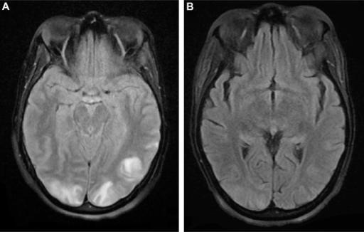
| current | 00:09, 4 February 2022 | | 512 × 326 (145 KB) | Ozzie10aaaa | Author:Chowdhary M, Kabbani AA, Tobey D, Hope TD, Department of Internal Medicine, Mercer University School of Medicine (Openi/National Library of Medicine) Source:https://openi.nlm.nih.gov/detailedresult?img=PMC4411014_ndt-11-1111Fig1&query=Posterior%20reversible%20encephalopathy%20syndrome&it=xg&req=4&npos=10 Description:f1-ndt-11-1111: MRI of the brain on admission revealing (A) cortical hyperintensity in the posterior parieto-occipital lobe, which suggests the presence of PRES; (B) follow... |
File usage
The following page uses this file: