File:PMC4370300 sfu14403.png
Jump to navigation
Jump to search
No higher resolution available.
PMC4370300_sfu14403.png (512 × 254 pixels, file size: 138 KB, MIME type: image/png)
File history
Click on a date/time to view the file as it appeared at that time.
| Date/Time | Thumbnail | Dimensions | User | Comment | |
|---|---|---|---|---|---|
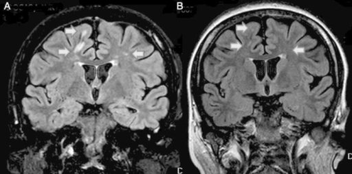
| current | 21:18, 29 October 2021 | | 512 × 254 (138 KB) | Ozzie10aaaa | Author:Ávila A, Vizcaíno B, Molina P, Gavela E, Perez-Ebri M, Pallardó L,Department of Nephrology , Hospital Universitario Dr Peset (Openi/National Library of Medicine) Source:https://openi.nlm.nih.gov/detailedresult?img=PMC4370300_sfu14403&query=Eculizumab&it=xg&req=4&npos=7 Description:SFU144F3: Evolution of MRI before (A) and after (B). (A) MRI before eculizumab therapy. High-intensity subcortical white matter lesions (arrow). Signs of mild cerebral atrophy. (B) MRI 3 weeks after eculizuma... |
File usage
The following page uses this file: