
No higher resolution available.
This file is from a shared repository and may be used by other projects.
The description on its file description page there is shown below.
License
Attribution 3.0 Unported (CC BY 3.0)
Summary
Author:Lehmann M, Barnes J, Ridgway GR, Ryan NS, Warrington EK, Crutch SJ, Fox NC, Dementia Research Centre, UCL Institute of Neurology, University College London (Openi/National Library of medicine) Source:https://openi.nlm.nih.gov/detailedresult?img=PMC4359276_figs1&query=Posterior%20cortical%20atrophy&it=xg&req=4&npos=2 Description:dfig1: Longitudinal changes in gray matter volume (sagittal view of the left hemisphere) in (A) posterior cortical atrophy (PCA) patients compared with control subjects and (B) typical amnestic Alzheimer’s disease (tAD) patients compared with control subjects. (C) Maps show regions that were most influential in making a classification between PCA and tAD groups. The color bar for the control comparisons (A and B) show t values for false discovery rate-corrected results (P < .05), with warmer colors indicating greater volume loss in PCA and tAD patients compared with control subjects. (C) Red represents areas where relatively lower gray matter volume change indicates PCA, whereas blue shows areas where lower gray matter volume change indicates tAD. A, anterior; P, posterior.
File history
Click on a date/time to view the file as it appeared at that time.
| Date/Time | Thumbnail | Dimensions | User | Comment |
|---|
| current | 16:46, 14 October 2021 | 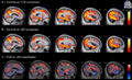 | 512 × 313 (234 KB) | Ozzie10aaaa | Author:Lehmann M, Barnes J, Ridgway GR, Ryan NS, Warrington EK, Crutch SJ, Fox NC, Dementia Research Centre, UCL Institute of Neurology, University College London (Openi/National Library of medicine) Source:https://openi.nlm.nih.gov/detailedresult?img=PMC4359276_figs1&query=Posterior%20cortical%20atrophy&it=xg&req=4&npos=2 Description:dfig1: Longitudinal changes in gray matter volume (sagittal view of the left hemisphere) in (A) posterior cortical atrophy (PCA) patients compared with control... |
File usage
The following page uses this file: