File:PMC4349084 fnhum-09-00108-g0002.png
Jump to navigation
Jump to search
No higher resolution available.
PMC4349084_fnhum-09-00108-g0002.png (512 × 198 pixels, file size: 106 KB, MIME type: image/png)
File history
Click on a date/time to view the file as it appeared at that time.
| Date/Time | Thumbnail | Dimensions | User | Comment | |
|---|---|---|---|---|---|
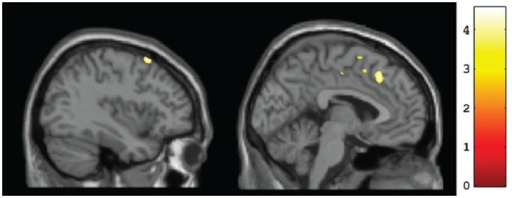
| current | 21:48, 21 October 2021 | 512 × 198 (106 KB) | Ozzie10aaaa | Author:Soh DW, Skocic J, Nash K, Stevens S, Turner GR, Rovet J,Department of Psychology, York University Toronto (Openi/National Library of Medicine) Source:https://openi.nlm.nih.gov/detailedresult?img=PMC4349084_fnhum-09-00108-g0002&query=Fetal%20alcohol%20syndrome&it=xg&req=4&npos=100 Description:F2: Sagittal views of regions showing significant (p <0.001, uncorrected) differences in gray matter volume at baseline for CT vs. FASD (TX and DTC combined): left superior frontal gyrus (BA8) [−37... |
File usage
The following page uses this file: