File:PMC4348355 40064 2015 883 Fig1 HTML.png
Jump to navigation
Jump to search
No higher resolution available.
PMC4348355_40064_2015_883_Fig1_HTML.png (512 × 281 pixels, file size: 122 KB, MIME type: image/png)
File history
Click on a date/time to view the file as it appeared at that time.
| Date/Time | Thumbnail | Dimensions | User | Comment | |
|---|---|---|---|---|---|
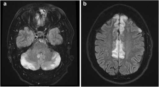
| current | 23:13, 5 February 2022 | | 512 × 281 (122 KB) | Ozzie10aaaa | Author:Edvardsson B,Department of Clinical Sciences, Lund University(Openi/National Library of Medicine) Source:https://openi.nlm.nih.gov/detailedresult?img=PMC4348355_40064_2015_883_Fig1_HTML&query=Hypertensive%20Encephalopathy&it=xg&req=4&npos=1 Description:Fig1: Primary MRI T2 and FLAIR images a and b displaying an increased signal in the cortex throughout the frontal and temporal lobes and in the cerebellum consistent with hypertensive encephalopathy/PRES. |
File usage
The following page uses this file: