File:PMC4303217 mgg30002-0472-f5.png
Jump to navigation
Jump to search
No higher resolution available.
PMC4303217_mgg30002-0472-f5.png (512 × 480 pixels, file size: 478 KB, MIME type: image/png)
File history
Click on a date/time to view the file as it appeared at that time.
| Date/Time | Thumbnail | Dimensions | User | Comment | |
|---|---|---|---|---|---|
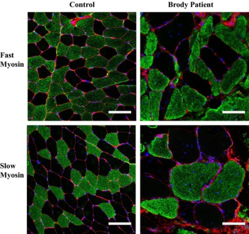
| current | 16:06, 19 October 2021 | | 512 × 480 (478 KB) | Ozzie10aaaa | Author:Sambuughin N, Zvaritch E, Kraeva N, Sizova O, Sivak E, Dickson K, Weglinski M, Capacchione J, Muldoon S, Riazi S, Hamilton S, Brandom B, MacLennan DH ,Defense and Veterans Center for Integrated Pain Management, Henry M. Jackson Foundation Rockville, Department of Anesthesiology, Uniformed Services University Bethesda (Openi/National Library of Medicine) Source:https://openi.nlm.nih.gov/detailedresult?img=PMC4303217_mgg30002-0472-f5&query=&req=4 Description:fig05: Immunofluorescence sta... |
File usage
The following page uses this file: