File:PMC4242703 ndt-10-2249Fig2.png
Jump to navigation
Jump to search

Size of this preview: 451 × 599 pixels. Other resolutions: 181 × 240 pixels | 512 × 680 pixels.
Original file (512 × 680 pixels, file size: 360 KB, MIME type: image/png)
File history
Click on a date/time to view the file as it appeared at that time.
| Date/Time | Thumbnail | Dimensions | User | Comment | |
|---|---|---|---|---|---|
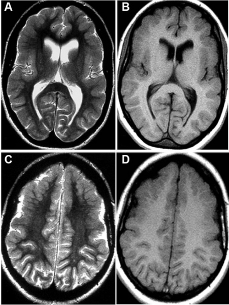
| current | 16:40, 21 January 2022 | | 512 × 680 (360 KB) | Ozzie10aaaa | Author:Conforti R, Capasso R, Capaldo G, Dato C, Saracino D, Di Iorio G, Melone MA,Neuroradiology Unit, Second University of Naples(Openi/National Library of Medicine) Source:https://openi.nlm.nih.gov/detailedresult?img=PMC4242703_ndt-10-2249Fig2&query=&req=4 Description:f2-ndt-10-2249: Axial Spin-Echo T2-weighted (A, C) and T1-weighted (B, D) magnetic resonance imaging study of the brain.Notes: Developmental abnormalities (polymicrogyria) in the frontal opercular cortex bilaterally, more evi... |
File usage
There are no pages that use this file.