File:PMC4241321 CRIPE2014-364657.002.png
Jump to navigation
Jump to search
No higher resolution available.
PMC4241321_CRIPE2014-364657.002.png (512 × 168 pixels, file size: 75 KB, MIME type: image/png)
File history
Click on a date/time to view the file as it appeared at that time.
| Date/Time | Thumbnail | Dimensions | User | Comment | |
|---|---|---|---|---|---|
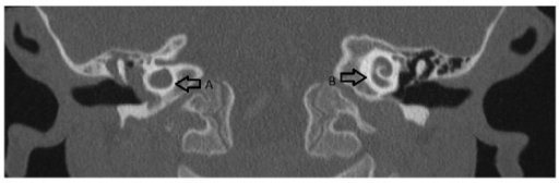
| current | 15:02, 26 December 2021 | 512 × 168 (75 KB) | Ozzie10aaaa | Author:Kepenekli-Kadayifci E, Karaaslan A, Atıcı S, Binnetoğlu A, Sarı M, Soysal A, Altınkanat G, Bakır M , Department of Pediatrics, Division of Pediatric Infectious Diseases, Pendik Training and Research Hospital, Marmara University Medical Faculty, Mimar Sinan Cad No. 41, Fevzi Cakmak Mah (Openi/National Library of Medicine) Source:https://openi.nlm.nih.gov/detailedresult?img=PMC4241321_CRIPE2014-364657.002&query=&req=4 Description:fig2: Computerized tomography of the temporal bone showin... |
File usage
There are no pages that use this file.