
No higher resolution available.
This file is from a shared repository and may be used by other projects.
The description on its file description page there is shown below.
License
Attribution 4.0 International (CC BY 4.0)
- &
CC0 1.0 Universal (CC0 1.0) Public Domain Dedication
Summary
Author:Jeong C, Lee JY, Kim J, Chae H, Park HI, Kim M, Kim OH, Kim P, Lee YK, Jung J,Catholic Genetic Laboratory Center, Seoul St, Mary' Hospital, The Catholic University of Korea(Openi/National Library of Medicine) Source:https://openi.nlm.nih.gov/detailedresult?img=PMC4236474_12891_2014_Article_2307_Fig1_HTML&query=Multiple%20epiphyseal%20dysplasia&it=xg&req=4&npos=3 Description:Fig1: Radiographs of proband at the age of 12 years. Anteroposterior view of knee joint showed shortening, fragmentation, and joint surface irregularity of distal femoral and proximal tibial epiphysis (A, B). Anteroposterior view of distal tibial epiphysis showed a lateral wedging and fragmentation (C, D). The tarsal navicular and cuneiform bone showed irregular ossification (E). The epiphysis of distal radius was wedge shaped and the epiphyses of the distal ulnae were relatively small and the carpal bone shows dysplasia and flattening (F). The epiphyses of proximal femur were spared (G).
File history
Click on a date/time to view the file as it appeared at that time.
| Date/Time | Thumbnail | Dimensions | User | Comment |
|---|
| current | 16:10, 2 August 2021 | 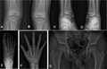 | 512 × 330 (133 KB) | Ozzie10aaaa | Author:Jeong C, Lee JY, Kim J, Chae H, Park HI, Kim M, Kim OH, Kim P, Lee YK, Jung J,Catholic Genetic Laboratory Center, Seoul St, Mary' Hospital, The Catholic University of Korea(Openi/National Library of Medicine) Source:https://openi.nlm.nih.gov/detailedresult?img=PMC4236474_12891_2014_Article_2307_Fig1_HTML&query=Multiple%20epiphyseal%20dysplasia&it=xg&req=4&npos=3 Description:Fig1: Radiographs of proband at the age of 12 years. Anteroposterior view of knee joint showed shortening, fragme... |
File usage
The following page uses this file: