No higher resolution available.
This file is from a shared repository and may be used by other projects.
The description on its file description page there is shown below.
License
Attribution 4.0 International (CC BY 4.0)
Summary
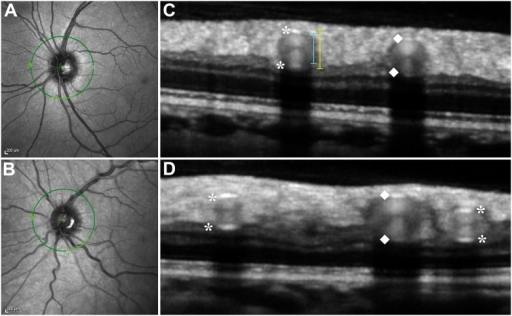
Author:Alten F, Motte J, Ewering C, Osada N, Clemens CR, Kadas EM, Eter N, Paul F, Marziniak M , Department of Ophthalmology, University of Muenster Medical Center(Openi/National Library of Medicine)Source:https://openi.nlm.nih.gov/detailedresult?img=PMC4221286_pone.0112311.g003&query=CADASIL&it=xg&req=4&npos=6 Description:pone-0112311-g003: A–D Combined simultaneous confocal scanning laser ophthalmoscopy (cSLO) and spectral-domain optical coherence tomography (SD-OCT).A–B Infrared cSLO image centered on the optic disc of a healthy control subject (A) and a CADASIL patient (B). Green circle indicates the position of corresponding SD-OCT scan. Light green section inferiorly on the circle marks the localization of corresponding SD-OCT scan shown aside. C–D Magnified SD-OCT scans of healthy control subject (C) and CADASIL patient (D) show sections of major retinal vessels appearing as a group of heterogeneous reflectivities in a round-shaped configuration. Asterisks mark the inner and outer reflections of arterial vessel walls and diamonds indicate inner and outer reflections of venous vessel walls. Hyperreflectivities representing the vessel walls seem thicker and more accentuated in the CADASIL patient. Particularly in veins, demarcation of the inferior vessel wall (towards the retinal pigment epithelium) often remains challenging due to absorption effects also seen as acoustical shadow underneath the vessel (towards the retinal pigment epithelium). Note the typical hour-glass shaped configuration within the vessel lumen in both subjects. Lateral vessel walls cannot be visualized as OCT laser beam is not projected perpendicularly to them.
File history
Click on a date/time to view the file as it appeared at that time.
| Date/Time | Thumbnail | Dimensions | User | Comment |
|---|
| current | 20:06, 19 February 2022 |  | 512 × 316 (166 KB) | Ozzie10aaaa | Author:Alten F, Motte J, Ewering C, Osada N, Clemens CR, Kadas EM, Eter N, Paul F, Marziniak M , Department of Ophthalmology, University of Muenster Medical Center(Openi/National Library of Medicine)Source:https://openi.nlm.nih.gov/detailedresult?img=PMC4221286_pone.0112311.g003&query=CADASIL&it=xg&req=4&npos=6 Description:pone-0112311-g003: A–D Combined simultaneous confocal scanning laser ophthalmoscopy (cSLO) and spectral-domain optical coherence tomography (SD-OCT).A–B Infrared cSLO image... |
File usage
There are no pages that use this file.