File:PMC3975201 cde-0006-0037-g03.png
Jump to navigation
Jump to search
No higher resolution available.
PMC3975201_cde-0006-0037-g03.png (512 × 309 pixels, file size: 340 KB, MIME type: image/png)
File history
Click on a date/time to view the file as it appeared at that time.
| Date/Time | Thumbnail | Dimensions | User | Comment | |
|---|---|---|---|---|---|
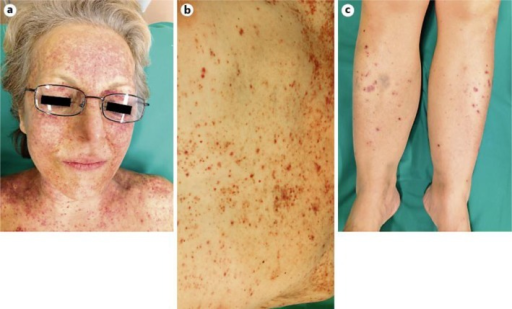
| current | 19:47, 11 August 2021 | | 512 × 309 (340 KB) | Ozzie10aaaa | Author:Kuonen F, Bucher M, de Leval L, Vernez M, Gilliet M, Conrad C, Feldmeyer L , Department of Dermatology and Venereology, Hôpital de Beaumont, Lausanne University Hospital Center(Openi/National Library of Medicine) Source:https://openi.nlm.nih.gov/detailedresult?img=PMC3975201_cde-0006-0037-g03&query=Hepatosplenic%20T-cell%20lymphoma&it=xg&req=4&npos=23 Description:F3: Photographs of the clinical evolution 2 months after the initial presentation. The clinical photographs show an intensif... |
File usage
There are no pages that use this file.