File:PMC3973996 1129-2377-15-13-3.png
Jump to navigation
Jump to search
No higher resolution available.
PMC3973996_1129-2377-15-13-3.png (512 × 491 pixels, file size: 236 KB, MIME type: image/png)
File history
Click on a date/time to view the file as it appeared at that time.
| Date/Time | Thumbnail | Dimensions | User | Comment | |
|---|---|---|---|---|---|
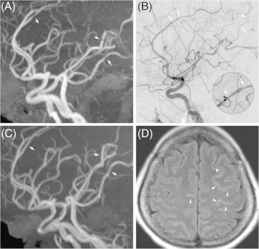
| current | 20:51, 31 January 2022 | | 512 × 491 (236 KB) | Ozzie10aaaa | Author:Cheng YC, Kuo KH, Lai TH,Section of Neurology, Department of Internal Medicine, Far Eastern Memorial Hospital(Openi/National Library of Medicine)Source:https://openi.nlm.nih.gov/detailedresult?img=PMC3973996_1129-2377-15-13-3&query=Reversible%20cerebral%20vasoconstriction%20syndrome&it=xg&req=4&npos=1 Description:F3: Imaging findings of reversible cerebral vasoconstriction syndrome. Multifocal vasoconstriction demonstrated by magnetic resonance angiography (MRA) (A) and catheter angiog... |
File usage
The following page uses this file: