File:PMC3965926 CRIOT2014-354672.002.png
Jump to navigation
Jump to search

Size of this preview: 396 × 599 pixels. Other resolutions: 159 × 240 pixels | 512 × 774 pixels.
Original file (512 × 774 pixels, file size: 507 KB, MIME type: image/png)
File history
Click on a date/time to view the file as it appeared at that time.
| Date/Time | Thumbnail | Dimensions | User | Comment | |
|---|---|---|---|---|---|
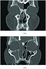
| current | 21:46, 2 March 2022 | | 512 × 774 (507 KB) | Ozzie10aaaa | Author:Hagiwara A, Nagai N, Ogawa Y, Suzuki M,Department of Otolaryngology, Tokyo Medical University, Department of Otolaryngology, Kohsei Chuo General Hospital(Openi/National Library of Medicine) Source:https://openi.nlm.nih.gov/detailedresult?img=PMC3965926_CRIOT2014-354672.002&query=&req=4 Description:fig2: CT showed an isodense mass which occupied the right nasal cavity, maxillary, ethmoid, and frontal sinuses, with no erosion of the bony walls. ↓: A small defect in the cribriform plate. |
File usage
The following page uses this file: