File:PMC3952252 er-3-2-70-5f3.png
Jump to navigation
Jump to search
PMC3952252_er-3-2-70-5f3.png (512 × 492 pixels, file size: 566 KB, MIME type: image/png)
Summary
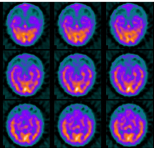
Author:Kang WH, Na JY, Kim MK, Yoo BG,Department of Neurology, Kosin University College of Medicine, Busan, Korea.( Openi US National Library of Medicine) Source:https://openi.nlm.nih.gov/detailedresult?img=PMC3952252_er-3-2-70-5f3&query=Hashimoto%27s%20Encephalopathy&it=xg&req=4&npos=10 Description:f3-er-3-2-70-5: Single photon emission computed tomography demonstrated decreased perfusion in the bilateral temporal regions.
Licensing
| This work is licensed under the Creative Commons Attribution-NonCommersial-ShareAlike 4.0 License. |
File history
Click on a date/time to view the file as it appeared at that time.
| Date/Time | Thumbnail | Dimensions | User | Comment | |
|---|---|---|---|---|---|
| current | 22:20, 28 July 2021 |  | 512 × 492 (566 KB) | Ozzie10aaaa (talk | contribs) | Author:Kang WH, Na JY, Kim MK, Yoo BG,Department of Neurology, Kosin University College of Medicine, Busan, Korea.( Openi US National Library of Medicine) Source:https://openi.nlm.nih.gov/detailedresult?img=PMC3952252_er-3-2-70-5f3&query=Hashimoto%27s%20Encephalopathy&it=xg&req=4&npos=10 Description:f3-er-3-2-70-5: Single photon emission computed tomography demonstrated decreased perfusion in the bilateral temporal regions. |
You cannot overwrite this file.
File usage
The following page uses this file:
