File:PMC3949000 kjp-52-85-g001.png
Jump to navigation
Jump to search
No higher resolution available.
PMC3949000_kjp-52-85-g001.png (512 × 121 pixels, file size: 170 KB, MIME type: image/png)
File history
Click on a date/time to view the file as it appeared at that time.
| Date/Time | Thumbnail | Dimensions | User | Comment | |
|---|---|---|---|---|---|
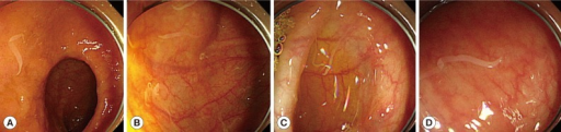
| current | 22:05, 16 December 2021 | 512 × 121 (170 KB) | Ozzie10aaaa | Author:Kim BJ, Song KS, Kong HH, Cha HJ, Ock M,Department of Internal Medicine, On Hospital(Openi/National Library of Medicine) Source:https://openi.nlm.nih.gov/detailedresult?img=PMC3949000_kjp-52-85-g001&query=Hymenolepiasis&it=xg&req=4&npos=9 Description:F1: Patient colonoscopy findings. Colonoscopy revealed that a large number of Hymenolepis nana adult worms were scattered throughout the colon as well as in the terminal ileum. (A) Terminal ileum. (B) Cecum. (C) Transverse colon. (D) Sigmo... |
File usage
The following page uses this file: