File:PMC3835646 kjr-14-963-g001 (1).png

From WikiProjectMed
Jump to navigation Jump to search

PMC3835646_kjr-14-963-g001_(1).png(512 × 288 pixels, file size: 38 KB, MIME type: image/png)

This file is from a shared repository and may be used by other projects. The description on its file description page there is shown below.

Summary

Description
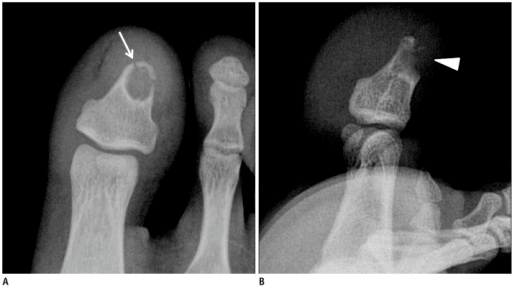
English⧼Colon⧽ F1: Fourteen-year-old male with desmoplastic fibroma in first toe.A, B. Fourteen-year-old male with intermittent painful swelling in his first toe. Anterior-posterior (A) and lateral (B) radiographs of forefoot demonstrate well-demarcated, sclerotic margined, oval, osteolytic lesion with dorsal cortical thinning and breakthrough (arrowhead) in distal phalanx of first toe. Associated pathologic fracture through distal portion of osteolytic lesion is demonstrated (arrow). There is no demonstrable matrix mineralization within tumor. C-F. MRI of desmoplastic fibroma of bone in first toe. Small ovoid central area of well-defined, osteolytic lesion shows hypointensity on all MR sequences (arrow in C-E), including axial T1WI (C), coronal T2WI (D), and sagittal STIR image (E), and shows little enhancement on sagittal fat-suppressed T1WI after gadolinium administration (F). Peripheral area of mass shows heterogeneous high signal intensity on T2WI and STIR images, and heterogeneous high to little contrast enhancement on fat-suppressed T1WI after gadolinium administration. Dorsal cortical breakthrough with small soft tissue extension (arrowheads in C, E, F) is demonstrated. T1WI = T1-weighted image, STIR = short tau inversion recovery. G, H. Photomicrographs of desmoplastic fibroma of bone. At low-power view (G), tumor displayed central hypocellular area (arrows) surrounded by peripheral cellular area (arrowheads) (hematoxylin and eosin stain, × 40). At high power view (H), peripheral area of tumor is composed of bland spindle cells with characteristic fascicular arrangement (arrows) (hematoxylin and eosin stain, × 200).
Date
Source https://openi.nlm.nih.gov/detailedresult?img=PMC3835646_kjr-14-963-g001&query=Desmoplastic%20fibroma&it=xg&req=4&npos=1
Author Kim OH, Kim SJ, Kim JY, Ryu JH, Choo HJ, Lee SJ, Lee IS, Suh KJ

Licensing

English⧼Colon⧽ This file is licensed CC BY-NC 3.0

This file was uploaded with UploadWizard.

File history

Click on a date/time to view the file as it appeared at that time.

Date/TimeThumbnailDimensionsUserComment
current22:28, 15 October 2022Thumbnail for version as of 22:28, 15 October 2022512 × 288 (38 KB)Ozzie10aaaaUploaded a work by Kim OH, Kim SJ, Kim JY, Ryu JH, Choo HJ, Lee SJ, Lee IS, Suh KJ from https://openi.nlm.nih.gov/detailedresult?img=PMC3835646_kjr-14-963-g001&query=Desmoplastic%20fibroma&it=xg&req=4&npos=1 with UploadWizard

There are no pages that use this file.