File:PMC3834874 1752-1947-7-255-1.png
Jump to navigation
Jump to search

Size of this preview: 491 × 599 pixels. Other resolutions: 197 × 240 pixels | 512 × 625 pixels.
Original file (512 × 625 pixels, file size: 209 KB, MIME type: image/png)
File history
Click on a date/time to view the file as it appeared at that time.
| Date/Time | Thumbnail | Dimensions | User | Comment | |
|---|---|---|---|---|---|
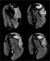
| current | 22:51, 15 January 2022 | | 512 × 625 (209 KB) | Ozzie10aaaa | Author:Kumar I, Verma A, Srivastava A, Shukla RC,Department of Radiodiagnosis and Imaging, Institute of Medical Sciences, Banaras Hindu University(Openi/National Library of Medicine) Source:https://openi.nlm.nih.gov/detailedresult?img=PMC3834874_1752-1947-7-255-1&query=Parsonage%20Turner%20syndrome&it=xg&req=4&npos=3 Description:F1: Magnetic resonance imaging of patient 1 at the level of shoulder girdle including the periscapular muscles. Sagittal T2-weighted fat-suppressed contiguous section... |
File usage
The following page uses this file: