File:PMC3806683 cop-0004-0099-g01.png
Jump to navigation
Jump to search

Size of this preview: 510 × 600 pixels. Other resolutions: 204 × 240 pixels | 512 × 602 pixels.
Original file (512 × 602 pixels, file size: 416 KB, MIME type: image/png)
File history
Click on a date/time to view the file as it appeared at that time.
| Date/Time | Thumbnail | Dimensions | User | Comment | |
|---|---|---|---|---|---|
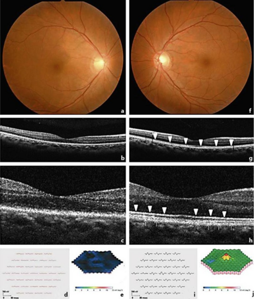
| current | 23:49, 13 November 2021 | | 512 × 602 (416 KB) | Ozzie10aaaa | Author:Makino S, Tampo H,Department of Ophthalmology, Jichi Medical University(Openi/National Library of Medicine) Source:https://openi.nlm.nih.gov/detailedresult?img=PMC3806683_cop-0004-0099-g01&query=Acute%20zonal%20occult%20outer%20retinopathy&it=xg&req=4&npos=7 Description:F1: Findings in a 39-year-old woman with AZOOR in the right (a–e) and left (f–j) eyes. Fundus photographs show no specific abnormalities (a, f). OCT horizontal scan (b) and high-magnification OCT findings (c) show an ab... |
File usage
There are no pages that use this file.