File:PMC3654851 mv-v19-935-f4 (1).png
Jump to navigation
Jump to search
No higher resolution available.
PMC3654851_mv-v19-935-f4_(1).png (512 × 282 pixels, file size: 243 KB, MIME type: image/png)
.png)
File history
Click on a date/time to view the file as it appeared at that time.
| Date/Time | Thumbnail | Dimensions | User | Comment | |
|---|---|---|---|---|---|
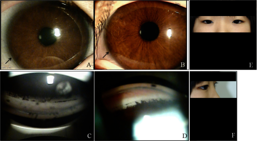
| current | 17:55, 24 September 2021 | | 512 × 282 (243 KB) | Ozzie10aaaa | Author:Kim GN, Ki CS, Seo SW, Yoo JM, Han YS, Chung IY, Park JM, Kim SJ ,Department of Ophthalmology, Gyeongsang National University, College of Medicine (Openi/National Library of Medicine) Source:https://openi.nlm.nih.gov/detailedresult?img=PMC3654851_mv-v19-935-f4&query=Axenfeld%E2%80%93Rieger%20syndrome&it=xg&req=4&npos=20 Description:f4: Ocular characteristics and systemic anomalies of patient II:2. Slit lamp photographs showed iris hypoplasia and posterior embryotoxon (black arrows) in... |
File usage
The following page uses this file:
.png)