File:PMC3615837 opth-7-561Fig3.png
Jump to navigation
Jump to search
Size of this preview: 445 × 599 pixels. Other resolutions: 178 × 240 pixels | 512 × 689 pixels.
Original file (512 × 689 pixels, file size: 420 KB, MIME type: image/png)
File history
Click on a date/time to view the file as it appeared at that time.
| Date/Time | Thumbnail | Dimensions | User | Comment | |
|---|---|---|---|---|---|
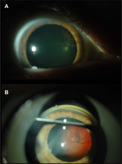
| current | 00:10, 2 February 2022 | | 512 × 689 (420 KB) | Ozzie10aaaa | Author:Sivaraman KR, Patel CG, Vajaranant TS, Aref AA,Department of Ophthalmology and Visual Sciences, Illinois Eye and Ear Infirmary, University of Illinois at Chicago School of Medicine(Openi/National Library of Medicine) Source:https://openi.nlm.nih.gov/detailedresult?img=PMC3615837_opth-7-561Fig3&query=&req=4 Description:f3-opth-7-561: Postdilation slit-lamp photo of the right eye of case 2 shows posterior pigment deposition on the posterior lens capsule (Scheie stripe), consistent with a... |
File usage
The following page uses this file: