File:PMC3612794 bmjopen2012002490f03 (1).png

From WikiProjectMed
Jump to navigation Jump to search

PMC3612794_bmjopen2012002490f03_(1).png(238 × 277 pixels, file size: 21 KB, MIME type: image/png)

This file is from a shared repository and may be used by other projects. The description on its file description page there is shown below.

Summary

Description
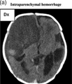
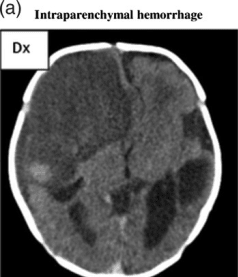
English⧼Colon⧽ BMJOPEN2012002490F3: (A) CT scan shows a very large intraparenchymal haemorrhage with mass effect and occupying most of the right frontal lobe in a term baby. Note a second but smaller haemorrhage on the left side adjacent to the ventricle but separate from this. The attenuation of both haemorrhages indicates that the haemorrhage is several weeks old at the time of imaging. We estimated the right haemorrhage to be 4–6 weeks old and the left still a couple of weeks older. The pregnancy was considered normal, and no intravenous immunoglobulin treatment was given. A live boy was born vaginally at 40 gestational weeks with petecchiae. The platelet count was 6×10⁹/l. (B) This CT shows marked ventriculomegaly, partly due to hydrocephalus, partly due to loss of brain tissue in the left hemisphere. Note the very large ventricle on the left side occupying most of the left hemicranium. The shape of the left lateral ventricle is irregular and that of residual from a previous intraventricular haemorrhage with a paraventricular atrophic defect caused by an old periventricular haemorrhagic infarction. This pattern is pathognomonic for this condition even though no actual blood can be detected. The interpretation of this image is that of an intraventricular haemorrhage associated with a periventricular haemorrhagic infarction timed at 28–30 gestational weeks or earlier. Hydrocephalus was diagnosed intrapartum because of breech presentation. A live boy was born at term with a platelet count of 4×10⁹/l. The child had severe cerebral palsy and a hydrocephalus shunt. (C) The CT scan shows multiple focal intraparenchymal haemorrhages throughout both cerebral hemispheres. Most haemorrhages are quite small. Note also the extensive extracranial subgaleal haemorrhage overlying the right hemicranium. All bleedings are of same age and maximum 7 days old. It was noted that the fetus was small on ultrasound scan at 25 weeks, but otherwise the pregnancy was considered normal. At 42 weeks a live boy was born vaginally, with multiple petecchiaes and multiple retinal bleedings. The platelet count at delivery was 14×109/l, with nadir value 8×10⁹/l. He received platelet transfusions. At time of discharge the boy was described as alive and well, but we do not have data on long-term clinical outcome.
Date
Source https://openi.nlm.nih.gov/detailedresult?img=PMC3612794_bmjopen2012002490f03&query=Intraparenchymal%20hemorrhage&it=xg&req=4&npos=4
Author Tiller H, Kamphuis MM, Flodmark O, Papadogiannakis N, David AL, Sainio S, Koskinen S, Javela K, Wikman AT, Kekomaki R, Kanhai HH, Oepkes D, Husebekk A, Westgren M

Licensing

{{subst:Custom license marker added by UW}} https://creativecommons.org/licenses/by-nc/2.0/ Attribution-NonCommercial 2.0 Generic (CC BY-NC 2.0)

This file was uploaded with UploadWizard.

File history

Click on a date/time to view the file as it appeared at that time.

Date/TimeThumbnailDimensionsUserComment
current19:48, 24 July 2022Thumbnail for version as of 19:48, 24 July 2022238 × 277 (21 KB)Ozzie10aaaaUploaded a work by Tiller H, Kamphuis MM, Flodmark O, Papadogiannakis N, David AL, Sainio S, Koskinen S, Javela K, Wikman AT, Kekomaki R, Kanhai HH, Oepkes D, Husebekk A, Westgren M from https://openi.nlm.nih.gov/detailedresult?img=PMC3612794_bmjopen2012002490f03&query=Intraparenchymal%20hemorrhage&it=xg&req=4&npos=4 with UploadWizard

There are no pages that use this file.