File:PMC3537700 1752-1947-6-281-3.png
Jump to navigation
Jump to search
No higher resolution available.
PMC3537700_1752-1947-6-281-3.png (512 × 384 pixels, file size: 538 KB, MIME type: image/png)
File history
Click on a date/time to view the file as it appeared at that time.
| Date/Time | Thumbnail | Dimensions | User | Comment | |
|---|---|---|---|---|---|
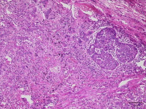
| current | 16:43, 22 November 2022 | | 512 × 384 (538 KB) | Ozzie10aaaa | Uploaded a work by Arai H, Inui K, Hashimoto K, Kan-O K, Nishii T, Kishida H, Okudela K, Tsuboi M, Nozawa A, Kaneko T, Masuda M from https://openi.nlm.nih.gov/detailedresult?img=PMC3537700_1752-1947-6-281-3&query=&req=4 with UploadWizard |
File usage
The following page uses this file: