No higher resolution available.
This file is from a shared repository and may be used by other projects.
The description on its file description page there is shown below.
License
Attribution 2.0 Generic (CC BY 2.0)
Summary
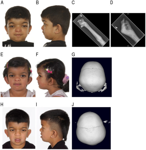
Author:Bendon CL, Fenwick AL, Hurst JA, Nürnberg G, Nürnberg P, Wall SA, Wilkie AO, Johnson D ,Oxford Craniofacial Unit, Oxford University Hospitals NHS Trust, John Radcliffe Hospital (Openi/National Library of Medicine) Source:https://openi.nlm.nih.gov/detailedresult?img=PMC3532175_1471-2350-13-104-1&query=Chromosome%205q%20deletion%20syndrome&it=xg&req=4&npos=30 Description:F1: A-D: Patient 1.A: Antero-posterior (AP) view showing facial features including hypertelorism. B: Lateral view showing brachycephaly and micrognathia. C: X-ray (XR) right radius/ulna showing broad appearance of the radius at the junction between the proximal and middle thirds. D: XR left hand showing crowding of the carpal bones, broad metacarpals, proximal and middle phalanges, and flexion at the MCP and PIP joints. E-G: Patient 2.E: AP view showing facial features including hypertelorism. F: Lateral view showing brachycephaly. G: 3D CT scan showing absence of the sagittal suture. H-J: Patient 3.H: AP view showing facial features including hypertelorism. I: Lateral view showing class III malocclusion and brachycephaly. J: 3D CT scan showing absence of the sagittal suture.
File history
Click on a date/time to view the file as it appeared at that time.
| Date/Time | Thumbnail | Dimensions | User | Comment |
|---|
| current | 20:49, 25 September 2021 |  | 512 × 524 (269 KB) | Ozzie10aaaa | Author:Bendon CL, Fenwick AL, Hurst JA, Nürnberg G, Nürnberg P, Wall SA, Wilkie AO, Johnson D ,Oxford Craniofacial Unit, Oxford University Hospitals NHS Trust, John Radcliffe Hospital (Openi/National Library of Medicine) Source:F1: A-D: Patient 1.A: Antero-posterior (AP) view showing facial features including hypertelorism. B: Lateral view showing brachycephaly and micrognathia. C: X-ray (XR) right radius/ulna showing broad appearance of the radius at the junction between the proximal and mid... |
File usage
There are no pages that use this file.