File:PMC3448114 cop-0003-0096-g01.png

From WikiProjectMed
Jump to navigation Jump to search

PMC3448114_cop-0003-0096-g01.png(512 × 464 pixels, file size: 308 KB, MIME type: image/png)

This file is from a shared repository and may be used by other projects. The description on its file description page there is shown below.

Summary

Description
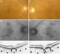
English⧼Colon⧽ F1: Findings in a 58-year-old man with bilateral focal choroidal excavation. a, c, e Images of the right eye. b, d, f Images of the left eye. a, b Fundus photographs on the initial presentation show RPE alterations beneath and adjacent to the fovea. c, d FAF image shows a focal area of hypoautofluorescence. e, f SD-OCT image with enhanced depth imaging through the fovea shows two concave-shaped conforming focal choroidal excavation (arrows) in both eyes. The excavations involved the outer retinal layers up to the ELM. The ELM band and the photoreceptor inner segment/outer segment junction can be seen at the excavation.
Date
Source https://openi.nlm.nih.gov/detailedresult?img=PMC3448114_cop-0003-0096-g01&query=Focal%20choroidal%20excavation&it=xg&req=4&npos=2
Author Katome T, Mitamura Y, Hotta F, Niki M, Naito T

Licensing

This work is licensed under the Creative Commons Attribution 3.0 unported License.

Anyone may use this image for any purpose provided it is attributed as specified.


&

Karger original article

This file was uploaded with UploadWizard.

File history

Click on a date/time to view the file as it appeared at that time.

Date/TimeThumbnailDimensionsUserComment
current21:07, 22 August 2022Thumbnail for version as of 21:07, 22 August 2022512 × 464 (308 KB)Ozzie10aaaaUploaded a work by Katome T, Mitamura Y, Hotta F, Niki M, Naito T from https://openi.nlm.nih.gov/detailedresult?img=PMC3448114_cop-0003-0096-g01&query=Focal%20choroidal%20excavation&it=xg&req=4&npos=2 with UploadWizard

There are no pages that use this file.