File:PMC3377144 IJOrtho-46-321-g002.png
Jump to navigation
Jump to search
No higher resolution available.
PMC3377144_IJOrtho-46-321-g002.png (512 × 372 pixels, file size: 178 KB, MIME type: image/png)
File history
Click on a date/time to view the file as it appeared at that time.
| Date/Time | Thumbnail | Dimensions | User | Comment | |
|---|---|---|---|---|---|
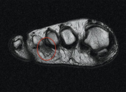
| current | 21:19, 14 October 2021 | | 512 × 372 (178 KB) | Ozzie10aaaa | Author:Torres-Claramunt R, Ginés A, Pidemunt G, Puig L, de Zabala S , Orthopaedic Department of the Parc de Salut Mar. Passeig Marítim (Openi/National Library of Medicine) Source:https://openi.nlm.nih.gov/detailedresult?img=PMC3377144_IJOrtho-46-321-g002&query=Morton%27s%20neuroma&it=xg&req=4&npos=3 Description:F2: Transverse T2-weighted MRI image shows low signal intensity mass in the same space. The Morton's neuroma is seen circled in red |
File usage
The following page uses this file: