File:PMC3307213 JCIS-2-5-g008.png
Jump to navigation
Jump to search

Size of this preview: 326 × 599 pixels. Other resolutions: 130 × 240 pixels | 504 × 926 pixels.
Original file (504 × 926 pixels, file size: 291 KB, MIME type: image/png)
File history
Click on a date/time to view the file as it appeared at that time.
| Date/Time | Thumbnail | Dimensions | User | Comment | |
|---|---|---|---|---|---|
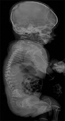
| current | 18:00, 13 December 2021 | | 504 × 926 (291 KB) | Ozzie10aaaa | Author:Kundaragi NG, Taori K, Jathar C, Disawal A,Department of Radiodiagnosis, Government Medical College(Openi/National Library of medicine) Source:https://openi.nlm.nih.gov/detailedresult?img=PMC3307213_JCIS-2-5-g008&query=Fibrochondrogenesis&it=xg&req=4&npos=8 Description:F7: Lateral radiograph of the spine. Severe platyspondyly with increased intervertebral disk spaces noted involving the entire vertebral column. Bodies of the vertebrae appeared small and pear-shaped on lateral view, wit... |
File usage
The following page uses this file: