File:PMC3305675 1532-429X-14-6-2.png

From WikiProjectMed
Jump to navigation Jump to search

PMC3305675_1532-429X-14-6-2.png(512 × 178 pixels, file size: 99 KB, MIME type: image/png)

This file is from a shared repository and may be used by other projects. The description on its file description page there is shown below.

Summary

Description
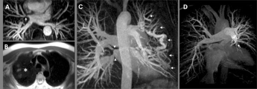
English⧼Colon⧽ F2: Abnormalities of Pulmonary Vasculature in Pulmonary Hypertension.; (A) White blood anatomy showing right upper pulmonary vein stenosis at the site of a prior ablation for atrial fibrillation (arrow). (B) Congestion and infarction in the right upper lobe on Half-Fourier Acquisition Single-Shot Turbo Spin-Echo images in the same patient (asterisk). (C) Magnetic Resonance Angiography from a separate patient with PH due to fibrosing mediastinitis; a varix is seen bypassing a stenosed left upper pulmonary vein (not shown) alongside stenoses of both right sided pulmonary veins (arrows). (D) Magnetic Resonance Angiography in patient with Chronic Thromboembolic Pulmonary Hypertension. The most striking feature is loss of the left descending pulmonary artery (arrow head).
Date
Source https://openi.nlm.nih.gov/detailedresult?img=PMC3305675_1532-429X-14-6-2&query=Pulmonary%20hypertension&it=xg&req=4&npos=11
Author Bradlow WM, Gibbs JS, Mohiaddin RH

Licensing

{{subst:Custom license marker added by UW}} https://creativecommons.org/licenses/by/2.0/ Attribution 2.0 Generic (CC BY 2.0)

This file was uploaded with UploadWizard.

File history

Click on a date/time to view the file as it appeared at that time.

Date/TimeThumbnailDimensionsUserComment
current20:03, 29 June 2022Thumbnail for version as of 20:03, 29 June 2022512 × 178 (99 KB)Ozzie10aaaaUploaded a work by Bradlow WM, Gibbs JS, Mohiaddin RH from https://openi.nlm.nih.gov/detailedresult?img=PMC3305675_1532-429X-14-6-2&query=Pulmonary%20hypertension&it=xg&req=4&npos=11 with UploadWizard

The following page uses this file: