File:PMC3290776 CDM-13-237 F9.png
Jump to navigation
Jump to search
No higher resolution available.
PMC3290776_CDM-13-237_F9.png (434 × 470 pixels, file size: 136 KB, MIME type: image/png)
File history
Click on a date/time to view the file as it appeared at that time.
| Date/Time | Thumbnail | Dimensions | User | Comment | |
|---|---|---|---|---|---|
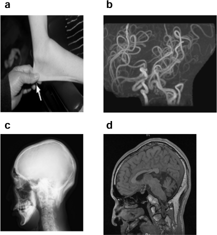
| current | 23:52, 16 February 2022 | | 434 × 470 (136 KB) | Ozzie10aaaa | Author:Kodama H, Fujisawa C, Bhadhprasit W,Department of health Dietetics, Teikyo Heisei University(Openi/National Library of Medicine) Source:https://openi.nlm.nih.gov/detailedresult?img=PMC3290776_CDM-13-237_F9&query=Occipital%20horn%20syndrome&it=xg&req=4&npos=1 Description:F9: a) Skin laxity in a 18 year-old patient with occipital horn syndrome.b) MRA showing tortuosity of cerebral arteries (arrow). c-d) Occipitalhorns are shown in a skull X-ray (c) and MRI T1 WI (d) (arrows). |
File usage
The following page uses this file: