File:PMC3261088 1750-1172-7-1-1.png
Jump to navigation
Jump to search
No higher resolution available.
PMC3261088_1750-1172-7-1-1.png (512 × 253 pixels, file size: 329 KB, MIME type: image/png)
File history
Click on a date/time to view the file as it appeared at that time.
| Date/Time | Thumbnail | Dimensions | User | Comment | |
|---|---|---|---|---|---|
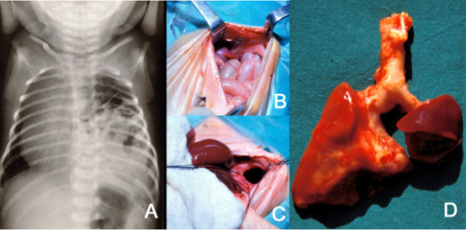
| current | 15:11, 4 November 2021 | | 512 × 253 (329 KB) | Ozzie10aaaa | Tovar,JA,Universidad Autonoma de Madrid, Department of Pediatric Surgery, Hospital Universitario La Paz (Openi/National Library of Medicine) Source:https://openi.nlm.nih.gov/detailedresult?img=PMC3261088_1750-1172-7-1-1&query=WAGR%20Syndrome&it=xg&req=4&npos=52 Description:F1: A: Plain X-ray of the thorax of a newborn with CDH. There are bowel loops into the left hemi-thorax, the mediastinum is displaced to the contralateral side and the space occupied by the lung is reduced. B and C: At lap... |
File usage
There are no pages that use this file.