File:PMC3229079 ad-23-S254-g002.png
Jump to navigation
Jump to search
No higher resolution available.
PMC3229079_ad-23-S254-g002.png (512 × 204 pixels, file size: 281 KB, MIME type: image/png)
File history
Click on a date/time to view the file as it appeared at that time.
| Date/Time | Thumbnail | Dimensions | User | Comment | |
|---|---|---|---|---|---|
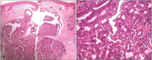
| current | 18:47, 11 April 2022 | 512 × 204 (281 KB) | Ozzie10aaaa | Author:Lee HJ, Lee D, Jung SY, Hong SK, Seo JK, Sung HS,Department of Dermatology, Busan Paik Hospital, Inje University College of Medicine(Openi/National Library of Medicine) Source:https://openi.nlm.nih.gov/detailedresult?img=PMC3229079_ad-23-S254-g002&query=Papillary%20hidradenoma&it=xg&req=4&npos=19 Description:F2: Variously shaped cystic and tubular structures were lined with a double or single layer of inner columnar and outer cuboidal cells. The cells of the lumina showed decapitation... |
File usage
There are no pages that use this file.