File:PMC2958498 IJHT2010-964513.002.png
Jump to navigation
Jump to search
No higher resolution available.
PMC2958498_IJHT2010-964513.002.png (478 × 339 pixels, file size: 190 KB, MIME type: image/png)
File history
Click on a date/time to view the file as it appeared at that time.
| Date/Time | Thumbnail | Dimensions | User | Comment | |
|---|---|---|---|---|---|
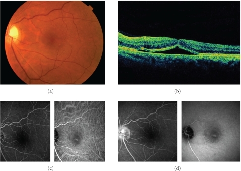
| current | 00:45, 26 February 2022 | | 478 × 339 (190 KB) | Ozzie10aaaa | Author:Hirano Y, Yasukawa T, Ogura Y, Department of Ophthalmology & Visual Science, Nagoya City University Graduate School of Medical Sciences (Openi/National Library of medicine) Source:https://openi.nlm.nih.gov/detailedresult?img=PMC2958498_IJHT2010-964513.002&query=retinal%20detachment&it=xg&req=4&npos=4 Description:fig2: The left eye of the same patient at the first visit. (a) Fundus photograph shows serous retinal detachment. The optic disc is swelling. (b) OCT shows retinal detachment i... |
File usage
The following page uses this file: