File:PMC2890894 jkms-25-1101-g003.png
Jump to navigation
Jump to search
No higher resolution available.
PMC2890894_jkms-25-1101-g003.png (512 × 185 pixels, file size: 256 KB, MIME type: image/png)
File history
Click on a date/time to view the file as it appeared at that time.
| Date/Time | Thumbnail | Dimensions | User | Comment | |
|---|---|---|---|---|---|
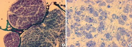
| current | 20:20, 21 December 2021 | 512 × 185 (256 KB) | Ozzie10aaaa | Author:Park YE, Yook JW, Kim DS, Department of Neurology, Pusan National University School of Medicine(Openi/National Library of Medicine) Source:https://openi.nlm.nih.gov/detailedresult?img=PMC2890894_jkms-25-1101-g003&query=&req=4 Description:F3: Pathological findings of right superficial peroneal nerve. (A) A profound loss of myelinated fibers is seen in one fascicle, while they are relatively spared in others (modified Gomori-Trichome, ×40). (B) On semithin sections, marked nerve fiber hy... |
File usage
The following page uses this file: