No higher resolution available.
This file is from a shared repository and may be used by other projects.
The description on its file description page there is shown below.
License
Attribution-NonCommercial 3.0 Unported (CC BY-NC 3.0)
Summary
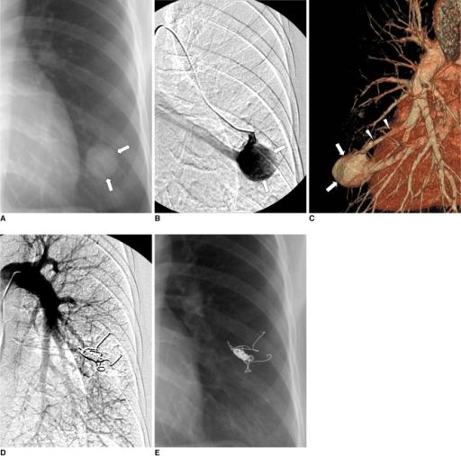
Author:Shin JH, Park SJ, Ko GY, Yoon HK, Gwon DI, Kim JH, Sung KB,Department of Radiology and Research Institute of Radiology, University of Ulsan College of Medicine, Asan Medical Center(Openi/National Library of medicine) Source:https://openi.nlm.nih.gov/detailedresult?img=PMC2864858_kjr-11-312-g001&query=Pulmonary%20arteriovenous%20malformation&it=xg&req=4&npos=3 Description:F1: 40-year-old female with large pulmonary arteriovenous malformation in left lower lobe (Patient No. 6). Simple radiograph (A), left selective pulmonary arteriogram (B), and CT angiogram (C) show large 32-mm-diameter pulmonary arteriovenous malformation (arrows) with single arterial feeder (arrowheads in C) and single draining vein. Left pulmonary arteriogram (D) after embolotherapy with placing four Nester coils shows no evidence of pulmonary arteriovenous malformation. Simple radiograph (E) 19 months after embolotherapy shows complete disappearance of pulmonary arteriovenous malformation shadow.
File history
Click on a date/time to view the file as it appeared at that time.
| Date/Time | Thumbnail | Dimensions | User | Comment |
|---|
| current | 21:26, 2 January 2022 |  | 512 × 507 (269 KB) | Ozzie10aaaa | Author:Shin JH, Park SJ, Ko GY, Yoon HK, Gwon DI, Kim JH, Sung KB,Department of Radiology and Research Institute of Radiology, University of Ulsan College of Medicine, Asan Medical Center(Openi/National Library of medicine) Source:https://openi.nlm.nih.gov/detailedresult?img=PMC2864858_kjr-11-312-g001&query=Pulmonary%20arteriovenous%20malformation&it=xg&req=4&npos=3 Description:F1: 40-year-old female with large pulmonary arteriovenous malformation in left lower lobe (Patient No. 6). Simple ra... |
File usage
The following page uses this file: