File:PMC2836895 JOP2009-203583.002.png
Jump to navigation
Jump to search
No higher resolution available.
PMC2836895_JOP2009-203583.002.png (419 × 195 pixels, file size: 151 KB, MIME type: image/png)
File history
Click on a date/time to view the file as it appeared at that time.
| Date/Time | Thumbnail | Dimensions | User | Comment | |
|---|---|---|---|---|---|
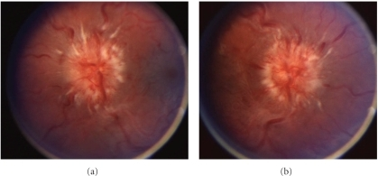
| current | 19:29, 1 January 2022 | | 419 × 195 (151 KB) | Ozzie10aaaa | Author:Bababeygy SR, Repka MX, Subramanian PS,Department of Ophthalmology, The Johns Hopkins Hospitals(Openi/National Library of Medicine) Source:https://openi.nlm.nih.gov/detailedresult?img=PMC2836895_JOP2009-203583.002&query=Papilledema&it=xg&req=4&npos=7 Description:fig2: Optic disc appearance, case 2. Fundoscopy of the left eye (a) and right eye (b) reveals grade IV papilledema as evidenced by severe elevation and hemorrhages. |
File usage
The following page uses this file: