File:PMC2667608 kjr-7-229-g003.png
Jump to navigation
Jump to search
No higher resolution available.
PMC2667608_kjr-7-229-g003.png (512 × 416 pixels, file size: 162 KB, MIME type: image/png)
File history
Click on a date/time to view the file as it appeared at that time.
| Date/Time | Thumbnail | Dimensions | User | Comment | |
|---|---|---|---|---|---|
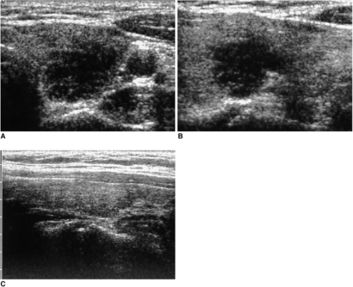
| current | 23:19, 18 January 2022 | | 512 × 416 (162 KB) | Ozzie10aaaa | Author:Park SY, Kim EK, Kim MJ, Kim BM, Oh KK, Hong SW, Park CS,Department of Diagnostic Radiology, Gachon University Gil Medical Center(Openi/National Library of Medicine) Source:https://openi.nlm.nih.gov/detailedresult?img=PMC2667608_kjr-7-229-g003&query=&req=4 Description:F3: A 50-year-old woman with neck swelling. Transverse (A) and longitudinal (B) sonograms of the left thyroid show an ill-defined, markedly hypoechoic lesion mimicking a malignant nodule. Subacute granulomatous thyroiditi... |
File usage
The following page uses this file: