File:PMC2374457 ar2378-1.png

From WikiProjectMed
Jump to navigation Jump to search

PMC2374457_ar2378-1.png(512 × 237 pixels, file size: 118 KB, MIME type: image/png)

This file is from a shared repository and may be used by other projects. The description on its file description page there is shown below.

Summary

Description
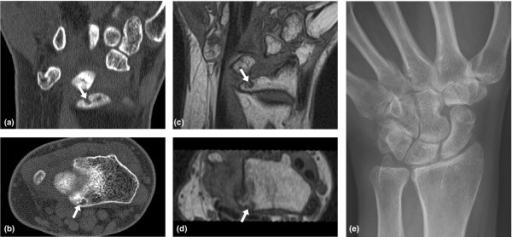
English⧼Colon⧽ F1: Erosions in the wrist of a rheumatoid arthritis patient. Wrist of a rheumatoid arthritis patient visualised by (a, b) computed tomography and (c, d) T1-weighted magnetic resonance imaging in the (a, c) coronal and (b, d) axial planes. A bone erosion at the distal radius is seen on both computed tomography and magnetic resonance images in two planes (white arrows), but not on the corresponding radiograph (e). The erosion was assigned an OMERACT erosion score of 1 on both computed tomography and magnetic resonance imaging.
Date
Source https://openi.nlm.nih.gov/detailedresult?img=PMC2374457_ar2378-1&query=Rheumatoid%20arthritis&it=xg&req=4&npos=15
Author Døhn UM, Ejbjerg BJ, Hasselquist M, Narvestad E, Møller J, Thomsen HS, Østergaard M

Licensing

{{subst:Custom license marker added by UW}} https://creativecommons.org/licenses/by/2.0/ Attribution 2.0 Generic (CC BY 2.0)

This file was uploaded with UploadWizard.

File history

Click on a date/time to view the file as it appeared at that time.

Date/TimeThumbnailDimensionsUserComment
current19:54, 23 September 2022Thumbnail for version as of 19:54, 23 September 2022512 × 237 (118 KB)Ozzie10aaaaUploaded a work by Døhn UM, Ejbjerg BJ, Hasselquist M, Narvestad E, Møller J, Thomsen HS, Østergaard M from https://openi.nlm.nih.gov/detailedresult?img=PMC2374457_ar2378-1&query=Rheumatoid%20arthritis&it=xg&req=4&npos=15 with UploadWizard

There are no pages that use this file.