File:PMC194618 1471-2369-4-6-1.png
Jump to navigation
Jump to search
No higher resolution available.
PMC194618_1471-2369-4-6-1.png (512 × 384 pixels, file size: 541 KB, MIME type: image/png)
File history
Click on a date/time to view the file as it appeared at that time.
| Date/Time | Thumbnail | Dimensions | User | Comment | |
|---|---|---|---|---|---|
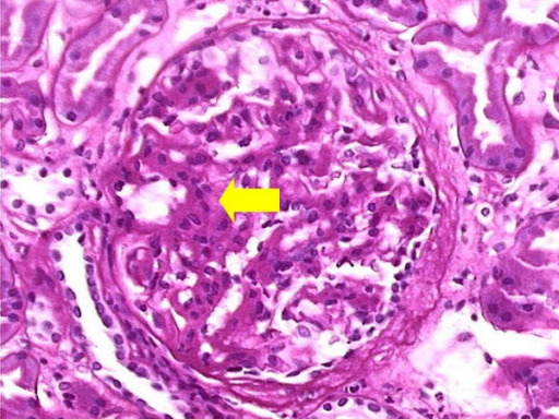
| current | 17:47, 4 January 2022 | | 512 × 384 (541 KB) | Ozzie10aaaa | Author:Fisher PW, Ho LT, Goldschmidt R, Semerdjian RJ, Rutecki GW,Department of Medicine, Northwestern University Feinberg School of Medicine, Evanston Northwestern Healthcare(Openi/National Library of Medicine) Source:https://openi.nlm.nih.gov/detailedresult?img=PMC194618_1471-2369-4-6-1&query=&req=4 Description:F1: Renal biopsy (Light Microscopy). Light microscopy image of a glomerulus demonstrating negative staining for both Congo-red and thioflavin-T. Expansion of the mesangium with incr... |
File usage
The following page uses this file: