File:PMC1562402 1746-160X-2-24-2.png
Jump to navigation
Jump to search
No higher resolution available.
PMC1562402_1746-160X-2-24-2.png (512 × 459 pixels, file size: 302 KB, MIME type: image/png)
File history
Click on a date/time to view the file as it appeared at that time.
| Date/Time | Thumbnail | Dimensions | User | Comment | |
|---|---|---|---|---|---|
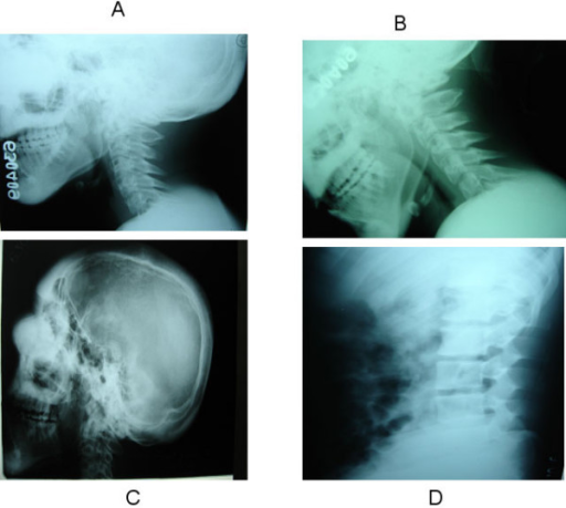
| current | 22:54, 16 January 2022 | | 512 × 459 (302 KB) | Ozzie10aaaa | Author:Maramattom BV ,Department of Neurology, Lourdes Hospital(Openi/National Library of Medicine) Source:https://openi.nlm.nih.gov/detailedresult?img=PMC1562402_1746-160X-2-24-2&query=Hyperostosis%20frontalis%20interna&it=xg&req=4&npos=10 Description:F2: Panel A and B show the cervical spine Xray in neutral position and flexion respectively. Note the hypertrophy of the posterior elements. Panel C shows diffuse cortical thickening on a lateral skull X-ray with soft tissue thickening and elev... |
File usage
There are no pages that use this file.