File:PMC1523325 1750-1172-1-22-1.png
Jump to navigation
Jump to search
No higher resolution available.
PMC1523325_1750-1172-1-22-1.png (512 × 355 pixels, file size: 330 KB, MIME type: image/png)
File history
Click on a date/time to view the file as it appeared at that time.
| Date/Time | Thumbnail | Dimensions | User | Comment | |
|---|---|---|---|---|---|
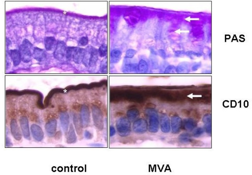
| current | 21:55, 23 January 2022 | | 512 × 355 (330 KB) | Ozzie10aaaa | Author:Ruemmele FM, Schmitz J, Goulet O,INSERM EMI 0212, Pediatric Gastroenterology, Hepatology and Nutrition, Hôpital Necker-Enfants Malades(Openi/National Library of Medicine)Source:https://openi.nlm.nih.gov/detailedresult?img=PMC1523325_1750-1172-1-22-1&query=Microvillus%20inclusion%20disease&it=xg&req=4&npos=1 Description:F1: High power magnification of a duodenal section of a patient with typical microvillous inclusion disease or microvillous atrophy (MVA) after periodic schiff acid (PAS... |
File usage
The following page uses this file: