File:PMC1386705 1476-7120-4-10-3.png
Jump to navigation
Jump to search
No higher resolution available.
PMC1386705_1476-7120-4-10-3.png (512 × 248 pixels, file size: 295 KB, MIME type: image/png)
File history
Click on a date/time to view the file as it appeared at that time.
| Date/Time | Thumbnail | Dimensions | User | Comment | |
|---|---|---|---|---|---|
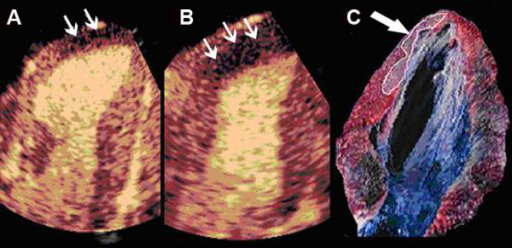
| current | 23:15, 8 October 2021 | | 512 × 248 (295 KB) | Ozzie10aaaa | Author:Dourado PM, Tsutsui JM, Chagas AC, Sbano JC, Aiello VD, Luz PL, Mathias W, Ramires JA, Heart Institute (InCor), University of São Paulo Medical School (Openi/National library of Medicine) Source:https://openi.nlm.nih.gov/detailedresult?img=PMC1386705_1476-7120-4-10-3&query=adenosine&it=xg&req=4&npos=4 Description:F3: Representative example of real-time myocardial contrast echocardiography images showing lack of perfusion that corresponds to infarcted area before (A) and during adenosin... |
File usage
The following page uses this file: