File:PDA Coil.png
Jump to navigation
Jump to search
No higher resolution available.
PDA_Coil.png (636 × 432 pixels, file size: 106 KB, MIME type: image/png)
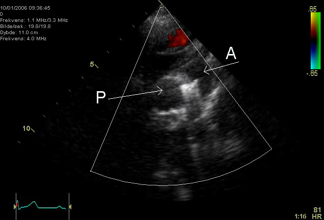
An echocardiogram of a coiled persisting ductus arteriosus. One can see the aortic arch,the pulmonary artery and the coil between them.
Image made by myself, Kjetil Lenes. Released in agreement with my employer.
| I, the copyright holder of this work, release this work into the public domain. This applies worldwide. In some countries this may not be legally possible; if so: I grant anyone the right to use this work for any purpose, without any conditions, unless such conditions are required by law. |
Keywords:
- en: Echocardiography, Sonography, Ultrasonography, Medical Ultrasound, Cardiology, persisting ductus arteriosus
- de: Echokardiographie, Sonografie, Sonographie, Ultraschall, Kardiologie, ductus arteriosus
File history
Click on a date/time to view the file as it appeared at that time.
| Date/Time | Thumbnail | Dimensions | User | Comment | |
|---|---|---|---|---|---|
| current | 16:50, 18 February 2006 | | 636 × 432 (106 KB) | commons>Ekko | An echocardiogram of a coiled persisting ductus arteriosus. One can see the aortic arch,the pulmonary artery and the coil between them. Image made by myself, Kjetil Lenes. Released in agreement with my employer. |
File usage
The following page uses this file: