File:Osteochondroma X-ray.jpg
Jump to navigation
Jump to search
Size of this preview: 382 × 599 pixels. Other resolutions: 153 × 240 pixels | 306 × 480 pixels | 952 × 1,494 pixels.
Original file (952 × 1,494 pixels, file size: 277 KB, MIME type: image/jpeg)
More images of this case:  
|
| Description |
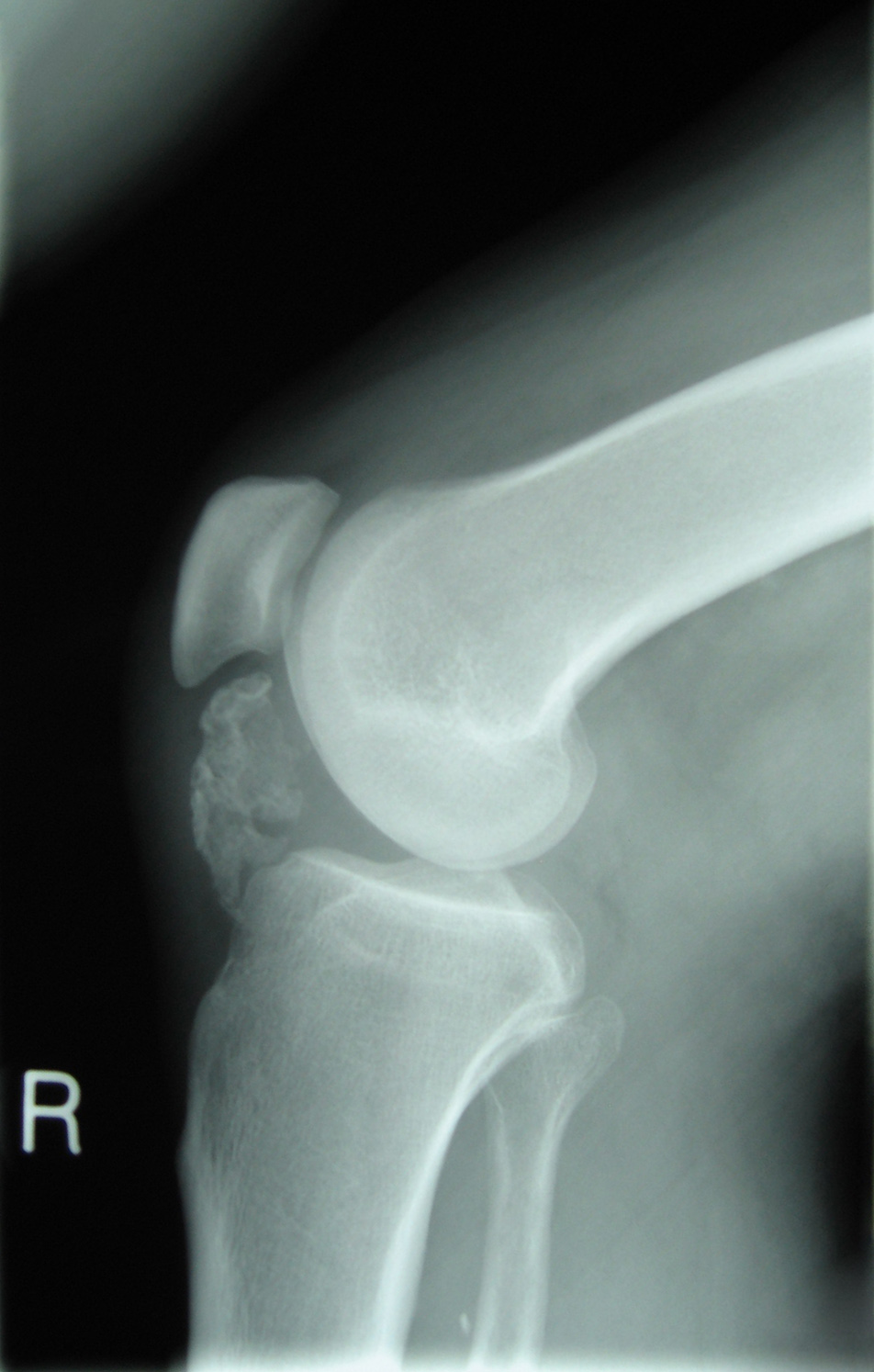
English: Lateral radiograph of the knee demonstrating ossification in the peritendinous tissues in a patient with osteochondroma. |
| Date | Published: 12 May 2008 |
| Source | Accelerated para-articular osteochondroma formation within the knee: a case report, Cases Journal. doi:10.1186/1757-1626-1-6 |
| Author | Michael R Carmont, Sian Davies, Daniel Gey van Pittius and Robin Rees |
| Permission (Reusing this file) |
This file is licensed under the Creative Commons Attribution 2.0 Generic license.
|
File history
Click on a date/time to view the file as it appeared at that time.
| Date/Time | Thumbnail | Dimensions | User | Comment | |
|---|---|---|---|---|---|
| current | 22:00, 17 July 2011 | | 952 × 1,494 (277 KB) | commons>Ras67 | perspective corrected and cropped |
File usage
There are no pages that use this file.